
The vascular tumours listed above are the commonest seen lesions. Below is a more comprehensive list with salient diagnostic features and links to virtual slides on Path Presenter. Many of the tips below are taken from Dirk Elston's book on Dermatopathology.
Angiokeratoma – bloody seb k! Acanthosis and large vascular superficial spaces
Nodular Kaposi’s sarcoma - usually dense spindle cell proliferation, plasma cells, mild atypia, blood, dorf balls! Vacuoles in vascular endothelium
Plaque stage Kaposi’s – slit like vascular spaces, plasma cells, wraps around adnexae and around itself, HHV8 stain
Glomus tumour – monotonous round evenly spaced cells around vascular space
Pyogenic granuloma ( lobular hemangioma ) – lobular vascular proliferation, sometimes a collarette, cf bacillary angiomatosis with lots of neutrophils
Angiolymphoid hyperplasia with eosinophilia – lymphoid hyperplasia plus a vascular proliferation and eosinophils, hob nailing of endothelium of larger vessels
Masson’s tumour Intravascular thrombus with intravascular proliferative hyperplasia
Angiosarcoma – Again slit like vascular spaces, hypercellular with more atypical endothelial cells
Lymphangioma – dilated spaces, may look like angiokeratoma, less overlying acanthosis
Bacillary Angiomatosis – Marked inflammation, blood, capillary vascular proliferation also necrosis and neutrophils, amphophilic bacterial colonies sometimes!
Arteriovenous malformation – mixture of sizes of vessels,
Spindle cell hemangioendothelioma – spindle cells, phleboliths
Targetoid hemosiderotic hemangioma – larger central vessel, slit like vascular spaces at periphery, hob nailing of endothelium and often hemosiderin, DD Kaposi’s with the vascular spaces
Lymphatic malformation - looks like a deeper lymphangioma
Targetoid Hobnail hemangioma – High vascular spaces, Staghorn vessels, promontory sign,
Microvenular hemangioma – mimics KS but HSV8 neg
Plaque KS – busy dermis, slit like spaces, patchy also around eccrine structures
Kaposiform hemangioma of Infancy – coagulation Kasaback Merric syndrome , looks like KS plaque type
Spindle cell hemangioma- Staghorn like spaces, spindle cell areas, Phleboliths, mimics nastier vascular tumours
Spindle cell hemangioendothelioma – behaves like low grade vascular tumour, Nodular and diffuse areas with lots of intracytoplastic lumens, Phleboliths
Pyogenic granuloma – capillary sized vessels, all arise from traumatic AV malformations., lobular with septae, erodes and can crust
Cherry angioma – vessels small but walls are thick and hyalinised
Angiosarcoma – dissecting crack like vessels, big hyperchromatic hobnail nuclei in vessels
Angiolymphoid hyperplasia with eosinophilia – AVM which becomes fancy, central thick vessel growing smaller vessels, with eosinophils
Bacillary angiomatosis – clusters of neutrophils deep with capillary proliferation in the dermis, amphophilic areas of organisms on silver stain, coccobaccili.
Intravascular papillary endothelial hyperplasia - Slit like vessels at edge of a clotted vessel, papillary projections
Glomangioma – thin layer of glomus cells, string of black pearls
Nodular Kaposi’s sarcoma – often no vascular spaces, red cells not in vascular spaces
Lymphangioma – spaces high with pink lymph
Eccrine angiomatous hamartoma – Amish tumour
Tufted hemangioma – plaque on shoulder of young child
Infantile hemangioma - Glut 1 pos ? embolic placenta!
Histiocytoid angiolymphoid hyperplasia with eosinophilia
Kaposi’s sarcoma – Patch stage , busy dermis, cells around eccrine glands,
Arteriovenous malformation (AVM) Thick and thin walled vessels, phleboliths
Angiokeratoma – high vascular spaces and overlying hyperkeratosis,
Epitheliod angioendotheliomatosis – lots of circles with intravascular lumens
Cavernous lymphangioma – deep cavernous lymph vessels, thin walled, may be staghorn like
Glomeruloid hemangioma – POEMS syndrome
Post mastectomy – angiosarcoma, Cmyc staining
KS and hobnail hemangioma can give hemosiderin around pre existing vessels
Angiosarcoma – can have KS like growth pattern but has high grade endothelial atypia
Verrucous hemangioma – verrucous surface
Glomus tumour
These lesions typically occur under the nail with a painful purple discolouration occurring on the nail bed, but they can also occur as solitary lesions elsewhere on the body. They are tumours of cells that control arterial venous anastomoses and basically are smooth muscle tumours. Histologically it is a well circumscribed and possibly encapsulated dermal tumour with just a few blood vessels surrounded by sheets of the glomus cells. These cells are quite round with round nuclei and sometimes eosinophilic cytoplasm. There is no variation in the cell size or shape. They are often described as being very monotonous. In a glomangioma usually you will get one or two layers of glomus cells around prominent vessels and usually glomangiomas are multiple. These vessels have thicker walls and they are probably a vascular malformation with a few glomus cells rather than glomus tumours which are actual tumours of glomus cells.
 |
This is the histology of a glomangioma with only a few layers of glomus cells around the vessels |


 |
Purplish sub ungual discoloration often painful of a glomus tumour |
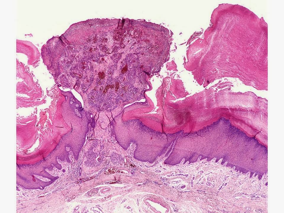
Pyogenic Granuloma
This tumour commonly arises in pregnancy at a site of minor injury. It can also occur on the gums where it is known as an epulis.It bleeds easily. Clinically it may have a smooth or eroded surface and a surrounding collarette of epidermis.
Histologically it is made up of lobules of closely packed endothelial cells. Mitoses may be seen.


Angiosarcoma
This tumour usually presents as a bruise like lesion on the head and neck of an elderly patient that is slowly growing. It can also present as a complication of chronic lymphedema on the legs. Histologically there are numerous blood vessels that are poorly demarcated. There are sometimes projections into the lumina and occasionally slit like spaces as much as you would see in a Kaposi's sarcoma. The endothelial cells are sometimes spindled or epitheliod and atypical and can vary between being mildly atypical to markedly atypical. There are often extravasated erythrocytes with hemosiderin staining and positive staining with endothelial cell markers such as CD31 or CD34.

 |
Note the prominent vascular spaces and the extravasated red cells |

Kaposi's sarcoma
 |
Nodule made up of spindled cells with slit like vascular spaces |


 |
Nodular Kaposi's sarcoma with spindled cells with erythrocytes between them and numerous slit like vascular spaces |
This used to be a rare vascular tumour seen in elderly Jewish males in Eastern Europe but with the advent of aids it became much commoner in a younger population. It is now known to be associated with the Herpes 8 virus and is a feature of early HIV and aids. It can present as bruise like macules or as papules and nodules, usually a purple colour, typically on the face and sometimes orally as well. It is essentially a vascular proliferation response to the Herpes 8 virus rather than a true sarcoma and it may begin in multiple areas at once. Typically histologically the dermis has slit like vascular spaces lined by spindled endothelial cells. Older lesions can have solid areas of spindle cells. Cellular atypia is usually mild but again there are often extravasated erythrocytes. Immuno staining for Herpes virus 8 is usually positive. The two major differentials are acroangiodermatitis, sometimes known a pseudo Kaposi's related to stasis. This condition may have some spongiosis and mature vessels and less of the slits. Secondly a pyogenic granuloma has a more rapid onset and more inflammation but again fewer slit like spaces.
 |
You might mistake this clinically for a melanoma |
 |
Dermatoscopy - The pigment is blood showing a parallel ridge pattern. |


No comments:
Post a Comment
Note: Only a member of this blog may post a comment.