Histologically the diagnosis of a Spitz nevus can be very difficult. The nests of melanocytes can be either epitheliod (like keratinocytes in the epidermis) or spindled but are often a mixture of both. The nests are usually high up in the epidermis explaining the dark colour of these lesions. Sometimes there is mild upward spread of individual melanocytes into the overlying epidermis but it is never extensive. The nests mature in depth as ordinary nevi do. There may be the occassional deeper mitosis but multiple mitoses are a sign to reassess the diagnosis. Sometimes the nests of cells in a Spitz nevus are said to hang or stream down from the rete rides like (hands of bananas!) The lesions are sharply defined laterally and there may be clefting around the nests. Kamino bodies which are pink accretions of the basement membrane are a prominent feature in some Spitz nevi. View the presentation below and then look at these Virtual Slides. Spitz NevusSpitz Nevus 2Intradermal Spitz NevusDesmoplastic Spitz
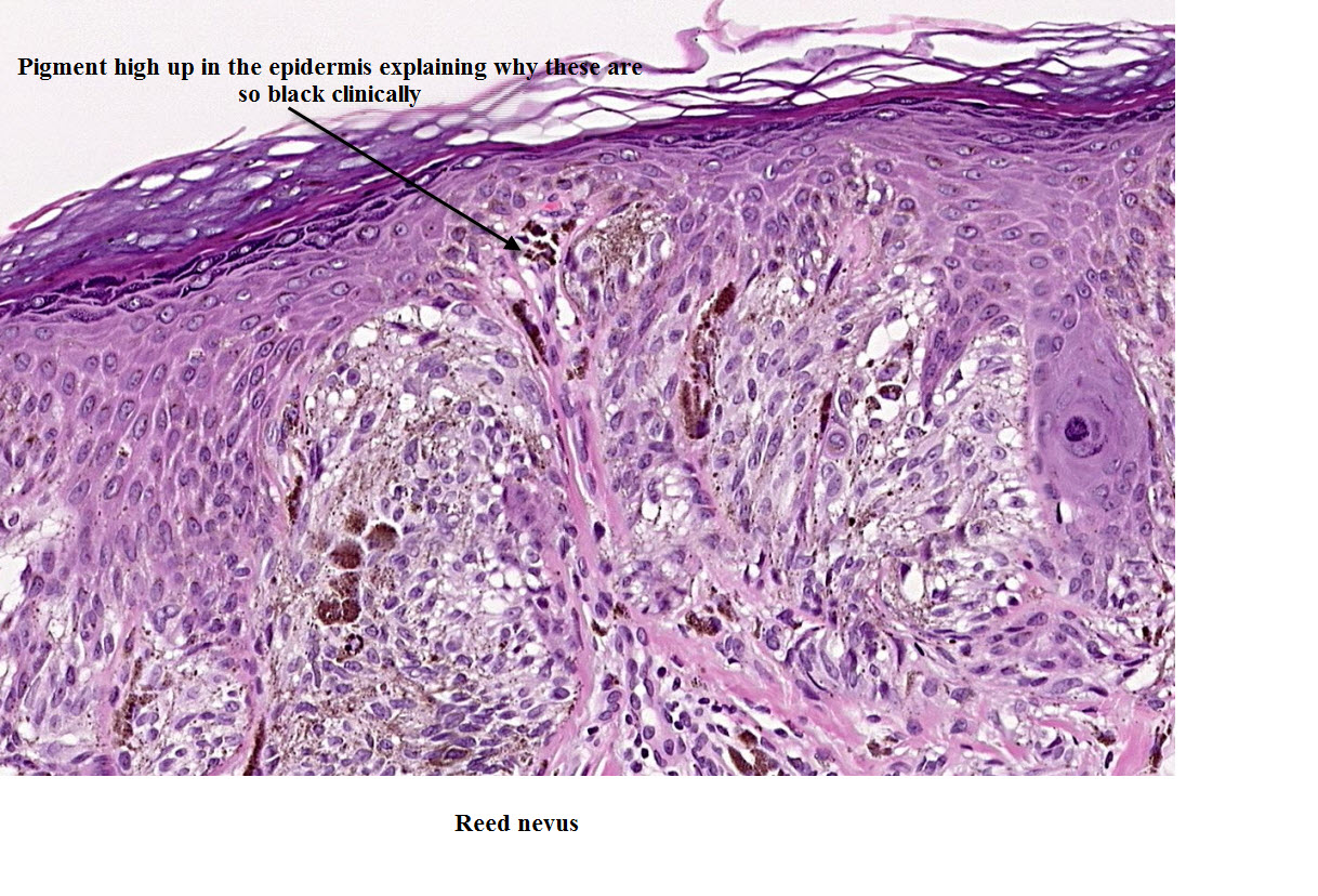
Reed Nevus This nevus is typically very dark with characteristic lines radial peripheral or streaks around the periphery under the dermatoscope. It is a worrying looking lesion clinically, often seen in younger individuals in their teens or twenties but is reasonably easy to diagnose under the microscope. It shows a lot of spindled junctional melanocytes often with the nests and cells orientated in a horizontal direction corresponding to the peripheral linear streaking.


No comments:
Post a Comment
Note: Only a member of this blog may post a comment.