These are tumours that you should just be able to recognise looking at a slide. The first one is Dermatofibrosarcoma Protuberans. These lesions are slow growing nodules that may occur at the site of trauma. They rarely metastasise and generally just grow locally. They often extend some distance beyond what is clinically apparent. The lesions presents as a dermal tumour as a nodule. There are usually uniform small spindle cells with plump nuclei and may be arranged in what is called a storiform pattern. They can infiltrate fat in a honeycomb pattern. There is usually thinning of the overlying epidermal rete ridges and you may get layers of fibrous tissue in the fat, that are aranged parallel to the overlying epidermis. A pigmented variant of DFSP is the Bednar tumour. In this the dendritic cells contain melanin and it may stain with S100. It particularly occurs in young women on the trunk and has never been known to metastasise. The melanin is probably due to colonisation of the DFSO bland melanocytes.
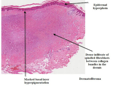
Dermatofibroma
Dermatofibroma
These lesions are easy to diagnose clinically but can be more difficult if you are just presented with a dermatoscopic view. They are thought to be a result of inflammatory changes in the skin and are said to be secondary to insect bite reactions.However recent molecular studies suggest they are true benign neoplasms.
Histologically there is increased dermal collagen arranged as whorls in an interlacing or storiform pattern.
Usually the overlying epidermis shows acanthosis with increased basal layer hyperpigmentation. Rarely basaloid hyperproliferation occurs giving a follicular or BCC like look to the skin overlying the dermal collagen bundles.
Various other cells can be found in the dermis including monster cells and Touton giant cells.
Shaved Dermatofibroma
Cellular Dermatofibroma
Aneurysmal Dermatofibroma
Hemosiderotic Dermatofibroma
Xanthomatous Dermatofibroma
Dermatofibroma with Monster Cells


No comments:
Post a Comment
Note: Only a member of this blog may post a comment.