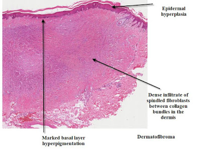
These lesions are easy to diagnose clinically but can be more difficult if you are just presented with a dermatoscopic view. They are thought to be a result of inflammatory changes in the skin and are said to be secondary to insect bite reactions.However recent molecular studies suggest they are true benign neoplasms.
Histologically there is increased dermal collagen arranged as whorls in an interlacing or storiform pattern.
Usually the overlying epidermis shows acanthosis with increased basal layer hyperpigmentation. Rarely basaloid hyperproliferation occurs giving a follicular or BCC like look to the skin overlying the dermal collagen bundles.
Various other cells can be found in the dermis including monster cells and Touton giant cells.
Shaved Dermatofibroma
Cellular Dermatofibroma
Aneurysmal Dermatofibroma
Hemosiderotic Dermatofibroma
Xanthomatous Dermatofibroma
Dermatofibroma with Monster Cells


No comments:
Post a Comment
Note: Only a member of this blog may post a comment.