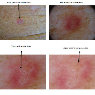
The Desmoplastic melanoma is a difficult melanoma to diagnose clinically. It can be a pink slightly keratotic nodule or a brown lentigo maligna like lesion. The latter presentation occurs because of an overlying proliferation of atypical melanocytes in the basal layer diagnosed as atypical melanocytic proliferation or a lentigo maligna. However in the deeper dermis there is a population of spindled melanocytes coursing between the collagen bundles and eliciting a desmoplastic response with marked thickening of the collagen. This causes the white lines seen dermatoscopically in some cases and also contributes to the white sclerotic areas seen clinically. Sometimes you also see localised aggregates of lymphoid cells in the dermis alerting you to the presence of the atypical spindled melanocytes. Some of these features are seen below. These tumours also show neurotropism which adds to their likelihood of being inadequately excised.
Look at the presentation below and then view the Virtual Slides
Desmoplastic Melanoma Desmoplastic Melanoma 2 Desmoplastic Melanoma 3 Scalp


No comments:
Post a Comment
Note: Only a member of this blog may post a comment.